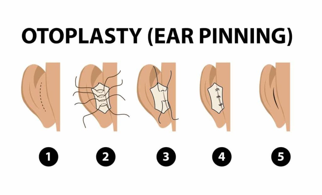
Otoplasty, also known as cosmetic ear surgery, is a procedure used to correct prominent ears or ears that are misshapen due to genetic defects or trauma. It can also be used to correct asymmetry between the two ears. The techniques used in otoplasty may vary depending on the type of corrections that need to be made, but typically involve reshaping or restructuring the cartilage of the ears and sometimes using sutures to secure them in the desired position.
The most common technique is called the “Mustarde” technique, which involves making an incision in the back of the ear and folding the cartilage to create a more aesthetically pleasing shape. Another technique, called the “Converse” technique, involves making incisions on the front and back of the ear and removing a small portion of the cartilage to produce the desired shape. In some cases, it may also be necessary to use sutures to secure the ear in the desired position in order to prevent it from reverting back to its original shape.
Table of Contents
ToggleIn this article we will discuss:
- Ear pinning
- Mustardé technique
- Stenström technique
- Anterior Scoring Technique in Otoplasty
- Final Words
- FAQ
Ear pinning – otoplasty techniques
Ear pinning is a type of otoplasty technique used to improve the appearance of protruding ears. This technique involves making an incision behind the ear and then folding the cartilage and suturing it in place. The surgeon may also remove excess skin or cartilage to achieve the desired results. The surgery is typically done under general anesthesia and the patient may be required to wear a headband or bandage for several days after the surgery to reduce swelling. Risks associated with ear pinning include bleeding, infection, scarring, numbness of the ear, and difficulty in fully closing the ear.

Mustardé technique
The Mustardé technique is a type of otoplasty, or ear surgery, that is used to correct prominent ears. It involves making an incision behind the ear and then suturing the cartilage in a way that creates a more natural-looking fold in the ear. The technique was developed by Dr. Paul Mustardé and has been used for many years to help patients achieve a more aesthetically pleasing appearance. This method, along with the Stenström and Converse methods, is a traditional form of otoplasty and is accomplished by suturing the antihelix.
The Mustardé technique of ear pinning is a surgical procedure where a long incision is made on the back of the ear and a strip of skin is removed. The cartilage between the helix and the sulcus posterior is then exposed and bent more strongly or formed anew with mattress sutures. The skin is then closed with sutures and a head bandage may be applied for up to two weeks. This technique leaves the cartilage fully intact and is not suitable for all ears.
There are potential complications associated with this procedure, such as bleeding, ear lying too close to the head, stronger asymmetry of the ear distances, hypertrophic scar, keloid, pressure damage (necrosis) caused by the bandage, and recurrence. A similar method, known as the Stitch method, is a closed and minimally invasive otoplasty, where the ear is not cut open.
Stenström technique – otoplasty techniques
Stenström first described the procedure for this surgery in 1963, and he made a small modification to it in 1973.
surgical procedure
This antihelix plastic surgery is performed using the scratch or scoring technique. This method is supported by the fact that cartilage, after being scratched or scored, bends convexly to the opposite side.
On the back of the ear, a lengthy incision is made, and a strip of skin is cut away. The skin and perichondrium are raised on the anterior surface of the antihelix through an incision in the cartilage of the cauda helicis (lower end of the ear cartilage) or the scapha.
The cartilage of the anterior (front side) of the antihelix is scored or scratched blindly using a rasp inserted into the skin-perichondrium tunnel that has formed. After suturing the skin wound on the back of the ear, a bandage is then placed on the area for one to two weeks, or longer in rare circumstances. For several days, ointment-containing swabs are used to model and support the cavum conchae, or scapha, on the front (anterior) side of the ear. Weerda believes that because of the limited elasticity of the cartilage, this method is not appropriate for older patients.
Anterior Scoring Technique in Otoplasty
The antihelical fold in otoplasty has long been produced by anterior scoring of the auricular cartilage. Many authors suggest using sutures in addition to subcutaneous cartilage scoring to create the antihelical fold with the least amount of access possible to the cartilage’s anterior surface.
For positioning the antihelical fold, other surgeons widely expose the anterior surface of the auricular cartilage and score it under close observation with or without sutures. The method we outline allows for a thorough exposure of the auricle’s anterior surface, weakening the cartilage and resulting in a smooth, uniform antihelical fold without the use of any long-term sutures. Additionally, it simultaneously corrects the concha’s height.
The cauda helicis is fixed behind the concha to control the position of the lobule, and the position of the helix is corrected by a subcutaneous section of the root of the helix to control the prominence of the upper third. Since every part of the auricle is accessible, it is a flexible technique that can be used to correct a variety of ear deformities.

Final Words about otoplasty techniques
Otoplasty, the surgical procedure used to correct protruding ears, typically combines incision, scoring, and suture techniques. The surgical approach is determined by the degree of the ear abnormality and the unique properties of the auricular cartilage. The incisions made in the ear during otoplasty are typically very small and are almost always hidden behind the ear, making it difficult to detect any scarring. Finally, some otoplasty procedures may also involve using silicon or other implants to further enhance the shape and size of the ear.

